Caused by the Plasmodium parasite, Malaria is a deadly disease that has plagued humanity for centuries. Microscopy’s contribution towards malaria detection began in the late 19th century when Sir Ronald Ross discovered the transmission of malaria by the Anopheles mosquito. This was a pivotal point in the journey of understanding Malaria- a breakthrough that enabled scientists to focus their efforts on developing accurate and efficient diagnostic methods, leading to the development of the first microscopy-based malaria diagnostic tests. From its humble beginnings to its current state-of-the-art applications, microscopy continues to be a vital tool in the world’s fight against malaria.
Better Diagnostics Through Latest Digital Technologies
There are quite a few drawbacks with traditional microscopy that impact its efficiency and accuracy such as its need for skilled microscopists/technicians for sample preparation and analysis, subjectivity in diagnosis, lack of digital records and deterioration of samples during transport and storage. There have been several technological advancements such as in molecular biology-based rapid diagnostics that have revolutionized the industry, but these methods can give only qualitative results and need to be followed by microscopy or PCR-based testing for confirmation and quantification.
Microscopy, thus, still remains a crucial component of malaria diagnosis, especially in regions that have limited resources and are unable to deploy expensive molecular tests. Digital microscopy, along with the integration of other advanced technologies such as AI and cloud computing is the answer to tackling most of the challenges associated with traditional microscopy. Skilled microscopists can identify and distinguish various species, P. falciparum, and P. vivax, being the most common, and determine the parasite load in a patient’s blood, aiding clinicians in choosing appropriate treatment regimens. However, with the recent advent of digital microscopes, the sample analysis process has the potential to become automated, standardized, more accurate and quicker with reduced scope for human error. Additionally, telemedicine and cloud-based technologies have enabled remote consultation and quality control. This means the turnaround time for diagnosis can be brought down from 1-2 weeks to a few hours, allowing for faster treatment and better recovery. Instead of collecting samples from peripheral settings and transporting them physically to referral centres for cross-verification, analysis and diagnosis can now be done online, thereby increasing efficiency. This has also facilitated collaboration between healthcare professionals and improved healthcare access in remote regions of the world.
The Future Of Malaria Microscopy
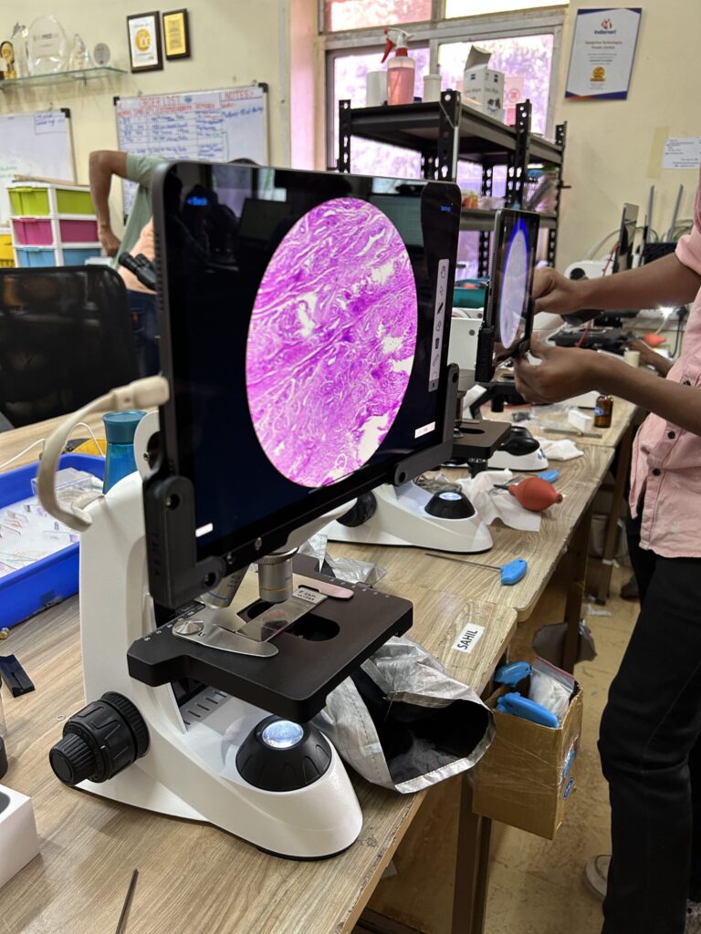
As technology continues to evolve, microscopy is poised to reach new heights in malaria detection. Researchers are exploring novel ways to enhance sensitivity, reduce processing time, and further automate the diagnostic process. Nanotechnology presents a promising avenue for revolutionizing microscopy in malaria detection. Nanoscale imaging platforms can potentially detect minute quantities of malaria parasites, enabling an even earlier diagnosis and timely intervention. These technologies hold particular significance in regions where asymptomatic carriers can unknowingly contribute to the transmission of the disease. Artificial intelligence and machine learning are being integrated into microscopy systems since AI-driven algorithms can analyze vast amounts of microscopy data rapidly, assisting in accurate parasite detection, species identification, and even predicting drug resistance patterns. For example, Medprime Technologies’ automated microscopy platform Micalys paired with an AI-based algorithm can not only detect malaria, but also identify the species, stage of infection and parasitic load. This IHF-Backed innovation has the potential to significantly improve the efficiency and accuracy of malaria diagnosis, making it more accessible and cost-effective. Beyond diagnosis, microscopy is also becoming instrumental in malaria research. High-resolution imaging techniques such as electron microscopy are enabling scientists to delve into the intricate interactions between the parasite and the human cells, leading to a better understanding of the disease’s mechanisms. This knowledge will significantly contribute towards the development of more effective drugs and vaccines against malaria.
Still A Long Way To Go
There are still multiple roadblocks in the widespread implementation of these newest tools such as AI-based malaria detection. Standardization of the sample preparation technique is a major challenge to be tackled. The use of various smearing and staining techniques by different labs and technicians can result in inconsistent results, leading to varying accuracies by automated malaria detection systems. Another issue is the commercial viability, especially in India. Malaria testing is done on a large scale mainly in the public health sector by national and state-level health programmes and the technology utilised has to be cost-effective for it to be affordable for the government to deploy on a large scale. This makes it unprofitable for private companies. While there are private companies that are actively working in this field currently, multi-sectoral collaboration along with funding and incentivization by the government, NGOs, funders, regulators, and other welfare organizations will go a long way in encouraging further R&D in this field.
By harnessing the power of microscopy and combining it with the benefits of other advanced technologies, we are inching closer to the dream of eradicating malaria from the face of the Earth.
About the Author:

Greeshma Unnikrishnan is the co-founder and Chief Operating Officer of Medprime Technologies. Having embarked on her entrepreneurial journey in 2014, she heads business development and fundraising for MedPrime Technologies. Under her leadership, the health-tech start-up has won several awards and grants for their innovations- Mumbai Entrepreneur Award 2022, Digital Trailblazer: NASSCOM 2022 and Magnetic Maharashtra Startup Awards 2018, to name a few. She holds an MTech degree from IIT Mumbai.